Dr Demetris Soteropoulos
Lecturer in Motor Systems Neuroscience
Telephone: +44 (0)191 208 6914
E-mail: demetris.soteropoulos@ncl.ac.uk
Anatomical Tracing
To help us understand the wiring of the nervous systen we also use anatomical tracing. This involves injecting a tracer in one part of the brain and then looking at another part to find evidence of the tracer. If the tracer is an anterograde tracer then it gets picked up by cells in the area of injection and gets transported elsewhere. A retrograde tracer gets picked up by presynaptic terminals at the site of injection and gets tranported back to the cell bodies wherever they may be. By using anterograde and retrograde tracers we can characterise the inputs and outputs of a given area.
We use well established and reliable tracers such as BDA and CtB (which are anterograde and retrograde tracers respectively), but have recently started using viral vectors as well as they offer some advantages over traditional ones. These include increased neuronal specificity depending on the protomers used as well as more complete dendritic labelling, which can be critical if one is looking at weaker connections that are likely to be located in more distal dendrites.
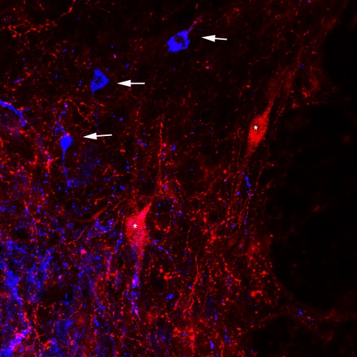
The image below shows an example of spinal interneurones in lamina VIII from the primate cervical spinal cord (data collected by Soteropoulos & Maxwell). In red are neurones (marked by '*') labeled with an AAV9 viral vector while in blue are cells (marked by arrows) labeled with CtB - note how much more extensive the dendritic labelling is for cells labeled with the viral vector compared to those with CtB.

More recently we have also started using dual vector systems that allow us to specifically target neurones that project from one area
to another, and in turn transiently silence those cells so that the role of the particular neural system can be studied.
Please see this publication from colleagues for further information.
Back to the techniques Page
Back to the main Lab Page

