Dr Demetris Soteropoulos
Lecturer in Motor Systems Neuroscience
Telephone: +44 (0)191 208 6914
E-mail: demetris.soteropoulos@ncl.ac.uk
Electrophysiology
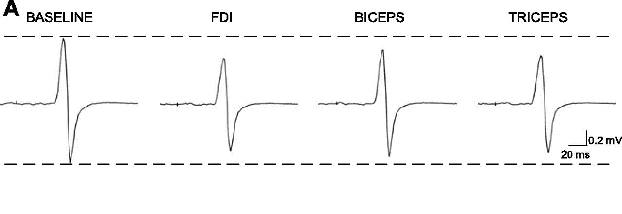
For our human experiments we record from the central and peripheral nervous system using non-invasive approaches. We can record from muscles and brain activity by using surface electrodes attached to the skin ( Electromyography and Electroencephalography respectively). Our lab is equipped with data capture systems and EEG/EMG amplifiers from CED and Digitimer
We are also able to non-invasively stimulate the central and peripheral nervous system. For cortical stimulation we primarily use Transcranial magnetic stimulation (TMS) as it a safe and non-noxious way to activate motor pathways.

For activation of peripheral nerves we use electrical stimulation which can allow us to activate specific classes of sensory fibres.
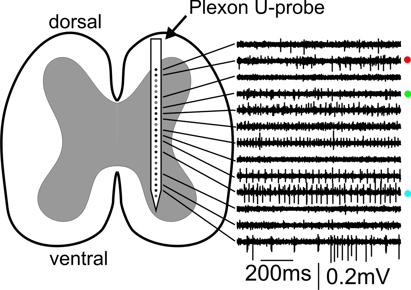
Current non-invasive approaches however are limited in the information they provide so we also carry out invasive recordings from animal models, either during behaviour and/or during terminal anaesthesia. These recordings consist primarily of local field potentials and neural spiking. To record these signals, we typically use multicontact probes from various sources (such as from Plexon, Thomas Recording, Neuronexus) and the data is captured using multichannel capture systems. The lab currently operates two 128Ch rigs. One consists of a 128ch TDT system allowing capture of up to 128Ch of spike and LFP/EMG data, as well as substantial online processing. In addition we also use a 128Ch system from Intan Technologies .

In addition to multichannel extracellular recordings, we also carry out intracellular recordings from motoneurones in the spinal cord, which allows us to directly measure synaptic inputs to these cells from various peripheral and central sources.
Neuronal Identification
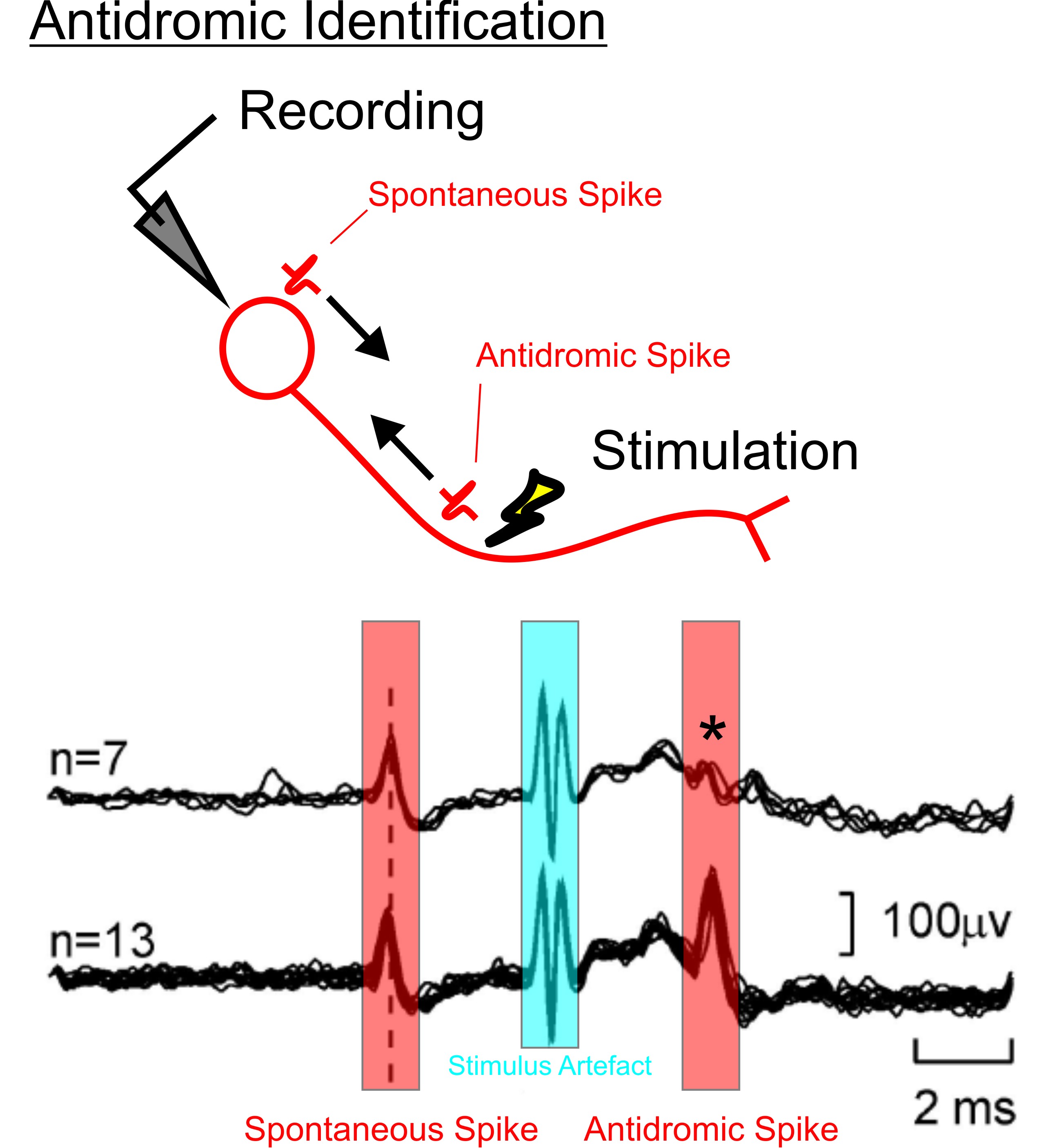
Not all neurones are the same in the brain and being able to record from anatomically separate neural populations allows much stronger conclusions to be made about how cell activity relates to movement. When recording neural activity, it is possible to identify projection neurones through the process of antidromic activation and collision testing.
If a stimulating electrode is placed in a tract through which a neurone sends an axon, then by stimulating that electrode an antidromic spike should be elicited that travels back towards the cell body. If the spontaneous spike is used to trigger stimulation at various delays, below a certain delay, the antidromic and orthodromic spikes should always collide, and after that delay they should never collide. This step provides verification that the spontaneously active cell used to trigger the stimulus does in fact send an axon down the tract of interest.

This is a technique that our lab (and the Motor Control Group in general) uses regularly, and this has allowed us to record from the activity of corticospinal cells, callosal cells, superior cerebellar peduncle cells (and others) during behaviour.
Back to the techniques Page
Back to the main Lab Page

